Glaucoma

Why Should I Bring my Pet to Willows for Treatment of Glaucoma?
Willows is one of Europe’s leading small animal referral centres. Our state-of-the-art hospital is led by internationally renowned Specialists, committed to providing the highest standards of veterinary care. Our team of Ophthalmic Specialists have a collective experience treating hundreds of patients with glaucoma. Our Ophthalmologists are supported by our multi-disciplinary team of Specialists across a number of disciplines including; Anaesthesia, Diagnostic Imaging and Emergency and Critical Care.
Willows also has a large dedicated team of Nurses and clinical support staff available 24 hours a day, every day of the year to provide the best possible care for your pet.
What is Glaucoma?
Glaucoma is an increased pressure inside the eye, caused by an obstruction to the drainage of the fluid from within the eye. In order to keep the eye inflated, fluid is normally produced and cleared away from the eye continuously.
What are the Most Common Causes of Glaucoma?
The blockage of fluid drainage within the eye can be due to an inherited defect (primary glaucoma) or due to another eye disease that interferes with drainage (secondary glaucoma).
Many cases of primary glaucoma are inherited and due to an abnormally formed drainage passage within the eye. Affected breeds include the Basset Hound, Welsh Springer Spaniel, Cocker Spaniel, Siberian Husky, Great Dane, the Flat Coated Retriever and many others. Often one eye is affected initially but there is a high risk that the other eye will follow at some point in the future. Common causes of secondary glaucoma are inflammation inside the eye or a shift of the position of the lens within the eye.
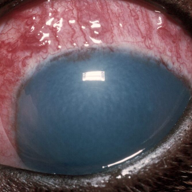
Fig 1: Acute glaucoma in a dog – note the blue discolouration of the cornea and severe inflammation
How is Glaucoma Diagnosed?
A special instrument called a tonometer (Tonovet or Tonopen) is used to measure the pressure inside the eye. A test (gonioscopy) is available to determine the predisposition of a patient to developing glaucoma. In this test, a special contact lens is applied to the eye to allow assessment of the structure concerned with drainage of fluid from the inside of the eye.
In most cases, the disease develops very rapidly. The patient is often depressed and reluctant to exercise. The eye becomes blind and appears red, painful and sore with a bluish tinge over the cornea. Less commonly, the pressure increase is slow and the clinical signs are not as noticeable. However, a gradual reduction in vision is often noticed.
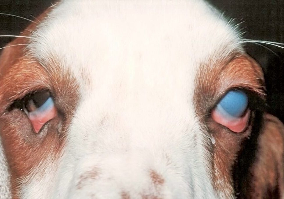
Fig 2: Acute gluacoma in the left eye – the cornea has become very opaque due to the high intraocular pressure
How can Glaucoma be Treated?
The aim of treatment in glaucoma is to preserve vision and to relieve the pain caused by the pressure increase. In order to reduce the pressure within the eye, drugs are given to reduce fluid production and to improve its removal from the eye. In some cases of secondary glaucoma, treatment of the underlying cause, such as anti-inflammatory medication or removal of a dislocated lens, can lead to the pressure decreasing.
Fig 3: A Tonovet is being used to measure the intraocular pressure

What can I Expect if my Pet is Treated for Glaucoma?
Patients with primary glaucoma are more difficult to treat and it is important to realise that no cure for the disease exists. In some patients, pressure control can be achieved with medical treatment only. However, with time, most patients becomes less responsive to the treatment and surgical alternatives may have to be considered. These include laser therapy or the surgical placement of a drainage implant into the affected eye. Regular check-ups will also be necessary to re-assess the second eye and check its pressure.
Eyes that have lost vision but continue to have an increased pressure are a cause of chronic pain for the patient. Removal of the eye (i.e. enucleation) must be considered in such cases to ensure the welfare and comfort of the patient. Occasionally, both eyes may unfortunately be lost. Should this be the case, most dogs will adapt very well to being blind and continue to lead a good quality life.
To save this page as a PDF, click the button and make sure “Save as PDF” is selected.
Ophthalmology
Find out more
To assist owners in understanding more about Ophthalmology we have put together a range of information sheets to talk you through the some of the more common conditions seen and treated by our Specialists.