Why should I bring my pet to Willows for an Indolent Ulcer?
Willows is one of Europe’s leading small animal referral centres. Our state-of-the-art hospital is led by internationally renowned Specialists, committed to providing the highest standards of veterinary care. As leaders in the field, our eye Specialists have experience of microsurgical techniques as well as access to a superb range of microsurgical instrumentation.
Our Ophthalmologists are supported by our multi-disciplinary team of Specialists across a number of disciplines including; Anaesthesia, Diagnostic Imaging and Emergency and Critical Care. Willows also has a large dedicated team of Nurses and clinical support staff available 24 hours a day, every day of the year to provide the best possible care for your pet.
What is the Cornea and what is an Indolent Ulcer?
The cornea is the clear window of the eye, a delicate structure, less than a millimetre thick. It consists of three layers:
- The epithelium ; the thin outer ‘skin’ of the cornea
- The stroma; the much thicker middle layer
- The endothelium; the very thin inner layer of the cornea (only one cell thick).
Any injury involving the cornea is known as an ulcer. Generally, corneal ulcers are described as superficial or deep. Ulcers that only involve the outer skin are called superficial ulcers or erosions. Ulcers that extend into the middle layer are known as deep ulcers. Most superficial ulcers heal rapidly as the cells of the surrounding outer ‘skin’ slide and grow into the defect. The new skin that grows then sticks to the tissue underneath. Most superficial ulcers will have healed within a week.
A Superficial Chronic Corneal Epithelial Defect (SCCED) or indolent ulcer is an ulcer which fails to heal in the expected time. It then tends to cause ongoing discomfort and irritation. Eyes affected with indolent ulcers try to grow a new surface skin over the defect, but the incoming cells fail to stick onto the middle layer. As a result, a thin layer of loose tissue can often be seen surrounding the ulcerated area. The reason why the cells fail to stick is not fully understood, however is believed that the epithelial cells fail to form tiny ‘feet’ that normally hold on to the tissue underneath.

What are the Signs of a Corneal Ulcer?
The cornea is sensitive due to the number of nerve endings, ulceration is usually associated with noticeable discomfort as the nerves are exposed. Signs of eye discomfort include weeping, blinking, squinting, pawing at the eye and general depression.
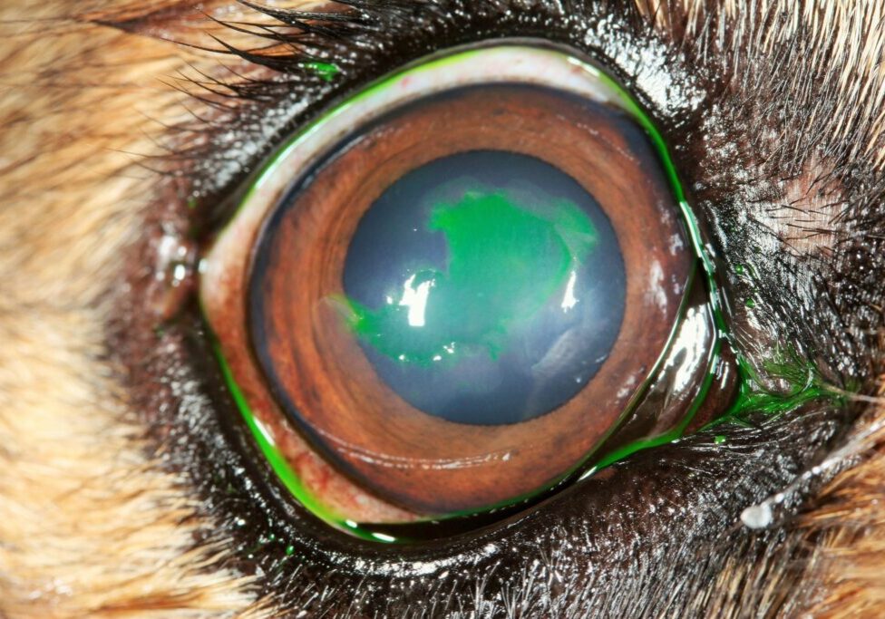
Fig 1: An indolent ulcer in a Boxer’s cornea
Certain breeds are predisposed to develop indolent ulcers including; Boxers, Corgis, Staffordshire Bull Terriers and West Highland White Terriers. However, any dog can develop an indolent ulcer, and older patients are more commonly affected. Once a dog has suffered an indolent ulcer in one eye, it may develop one in the other eye, or recurrence of ulceration in the first eye. This can happen at any time after the first ulcer and sometimes years later.


What Treatments are Available if my Dog has an Indolent Ulcer?
Indolent ulcers cannot be healed with topical medical treatment alone. In order for healing to take place, it is important that all loose tissue is removed and that the exposed middle layer is treated and ‘freshened up’ to allow adhesion of new ‘skin’ cells. Three main treatment options are available:
Debridement with cotton-tipped applicator: Approximately 50% of SCCEDs heal with this treatment (often performed without sedation)
Debridement of the lesion in conjunction with a diamond burr keratotomy: Approximately 85% of SCCEDs heal with this treatment within two weeks (often performed under sedation)
The option of surgery for an indolent ulcer may have to be reconsidered if it fails to heal after several attempts at debridement and/or diamond burr keratotomy. As such a superficial keratectomy (under general anaesthetic) may be discussed. Approximately 100% of SCCEDs heal with this treatment within ten days with the removal of all diseased skin and some underlying middle layer. Whilst the operation has a high success rate it is not always suitable for every patient.
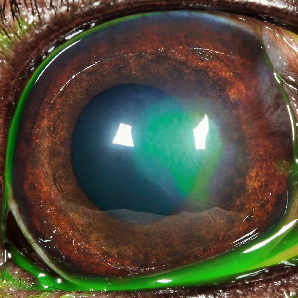
Fig 2: The same ulcer, now stained with a dye to show up the defect.
What can I Expect if my Dog is Treated for an Indolent Ulcer?
Usually, a broad spectrum antibiotic will be given to be applied three times a day to help to prevent infection. In some cases, it may be necessary to give a drug (atropine) which widens the pupil and reduces pain associated with the ulcer. Often patients with an indolent ulcer will receive a painkiller which is given with food.
A bandage contact lens can be placed in the eye at the end of the procedure. Such a lens is not likely to significantly affect the healing time but will cover exposed nerve endings and provide pain relief. In some patients, healing of the ulcer occurs fast and the cornea will only be slightly cloudy during treatment. However, in patients where healing is slow, it is common to find that blood vessels grow into the cornea and that pink granulation tissue forms to cover the defect. During this time, the eye may appear very red and odd looking. However, once the defect is fully covered, the granulation tissue will gradually clear over a period of months, leaving only minor corneal scarring in the majority of cases. Vision in most patients will return to normal or near normal after an episode of indolent ulceration.
Until healing of the indolent ulcer is achieved, it is advisable that the patient returns to the Specialist to ensure satisfactory progress. On occasions, treatment will be shared between your local Vet and the eye Specialist to reduce the travelling involved for you. Once the ulcer has fully healed, patients should not require frequent Veterinary attention and a return to the eye Specialist will only be necessary in selected cases or if the problem recurs in either eye.
To save this page as a PDF, click the button and make sure “Save as PDF” is selected.
Ophthalmology
Find out more
To assist owners in understanding more about Ophthalmology we have put together a range of information sheets to talk you through the some of the more common conditions seen and treated by our Specialists.

