Dogs with Twisted Stomachs
Why Should I Bring my Pet to Willows for a Gastric Dilation Volvulus?
Willows is one of Europe’s leading small animal referral centres. Our state-of-the-art hospital is led by internationally renowned Specialists, committed to providing the highest standards of veterinary care. At Willows our Specialist-led Emergency and Critical Care service is based within our advanced Intensive Care Unit and is fully supported by our experienced multi-disciplinary team including Diagnostic Imaging, Anaesthesia, Soft Tissue Surgery, Orthopaedics and Oncology.
All of which is supported by our dedicated team of Nurses and clinical support staff are available 24 hours a day, every day of the year to provide the best possible care for your pet.
What is Gastric Dilation Volvulus?
Gastric Dilatation and Volvulus (GDV) is a condition where the stomach bloats (dilatation), increases in size and then rotates (volvulus) around itself. It is an acute, life-threatening condition that needs urgent Veterinary intervention. If your dog at any time starts retching, has unproductive vomit, they should be seen as an emergency by a Veterinarian. In cases of GDV, this can be a life threatening condition which could be fatal if no emergency care is taken.
What are the Most Common Causes of GDV?
It has been reported in many breeds of dogs and is rarely seen in cats. The condition is not fully understood yet but it has been associated with certain risk factors:
- Large and giant-sized dog breeds and lean body condition
- Adult to geriatric age
- Consumption of a large meal once daily / being fed dry food only
- Eating quickly
- Heavy exercise after a meal
- Deep chest (increased thoracic height to width ratio)
- A stressful event
What are the Signs of a GDV in a Dog?
It is unclear if bloating or rotation occurs first in the development of GDV, however it is assumed that volvulus occurs first. Bloating of the stomach results from an accumulation of gas and/or fluid, and the rotation prevents the normal release of these contents. After rotation of the stomach, gas is trapped within this compartment and intragastric pressure rises. Splenic entrapment often accompanies GDV. The increasingly swollen stomach compromises venous return by compression of the caudal vena cava and the abdominal organs become deprived from perfusion. In addition, the stomach pushes the diaphragm forward, reducing the space that is available for the lungs. These changes cause more severe symptoms to appear, such as panting, weakness and even collapse due to shock.
The initial signs that can be observed in a pet with GDV are often associated with abdominal pain. These can include but are not limited to:
- Restlessness, refusing to sit down and relax
- Looking at the abdomen, anxious
- Drooling
- Distended/bloated and uncomfortable abdomen
- Retching without producing anything
- Collapse
- Difficulty breathing
How is GDV Diagnosed?
The history and a physical examination of the patient are very important and can raise the alarm of a possible GDV. The management of the patient then needs to be as fast as the condition progresses and it can often be challenging.
A combination of tests are needed to fully establish the status of the patient. A full blood profile, including biochemistry, complete blood count and electrolytes is important to check for metabolic imbalances, the degree of bleeding due to vessels distension but also other factors such as glucose and lactate. Blood gas analysis can also be performed to evaluate the nature and severity of the respiratory compromise and hypotension/hypoperfusion of organs.
Furthermore, the patient needs to be closely monitored with an electrocardiogram (ECG), in order to evaluate the presence of cardiac arrhythmias (irregular rhythm and rate of the heart) which are commonly seen later in the course of the disease. The condition is confirmed with abdominal X-rays of at least two different views.
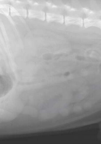
Fig 1: Normal shape and position of an empty stomach in the cranial part of the abdomen (black arrow)
What Treatments are Available for Dogs GDV?
During all tests, the patient will receive oxygen and strong pain relief via intravenous fluids. Any other abnormalities on the blood tests or on the ECG are treated accordingly to achieve stabilisation of the patient.
Once GDV is confirmed, gastric decompression follows, this includes the passing of a tube down the esophagus into the stomach to release the air and fluid accumulation and lavage (flushing of water) into and out of the stomach to remove remaining food particles. In cases of severe bloating, a needle or catheter may be placed into the stomach first, from the outside of the body to release air and aid in the passing of the tube and the tube could be placed during the general anesthesia just before surgery.
Decompression of the stomach will be a short term solution, the rotation in the stomach needs to be corrected. The only way to get the stomach back in the right position is by surgery. Surgery involves full exploration of the abdomen and de-rotation of the stomach. Additionally, the viability of the stomach wall, the spleen, and all other organs will be determined. Removal of part of the stomach wall (partial gastrectomy) or the spleen (splenectomy) is performed if deemed appropriate. Once the stomach is returned to the normal position, it needs to be permanently fixed to the abdominal wall (gastropexy) to prevent volvulus if subsequent gastric dilatation occurs again.
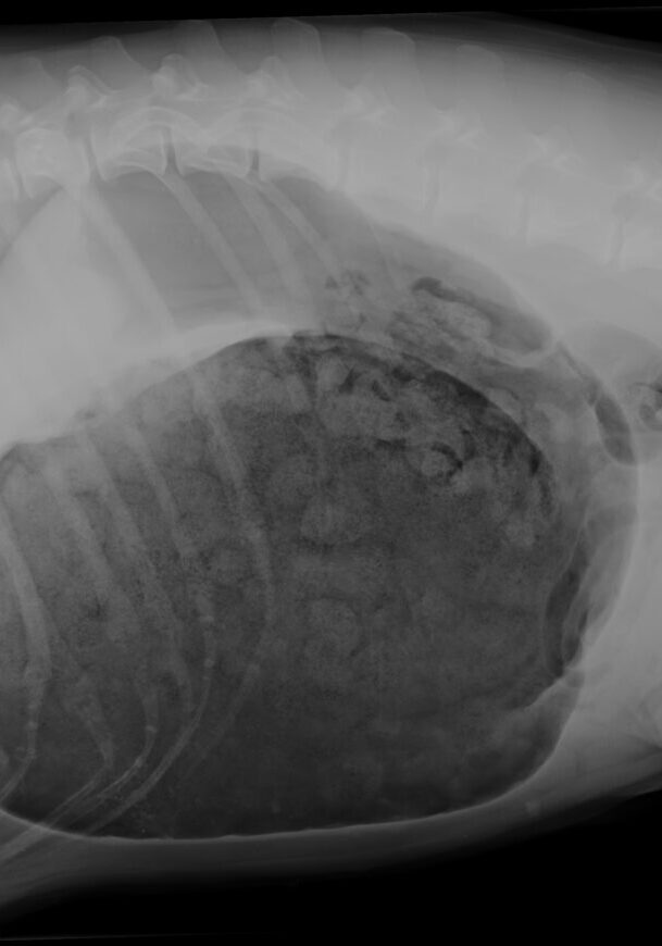
Fig 2: Severely distended, air and food filled twisted stomach. Abnormal position of the stomach with the “double bubble” image characteristic of GDV
Prognosis and Long Term Management
As gastric dilatation worsens and full body effects become prolonged, many secondary complications may occur such as poor oxygen delivery to many tissues. This can lead to cell death in the liver, kidneys, and other vital organs. Toxins may also increase locally, and when the stomach is deflated, may circulate through the body resulting in additional cardiac arrhythmias, acute kidney failure, and liver failure. Bacteria also commonly gain access to the blood during this condition leading to bacteremia (bacteria in the blood) and sepsis.
Mortality rates associated with GVA are approximately 15%. Mortality and morbidity (complication) rates increase as disease severity and time increase. Factors that have been shown to increase mortality rate include patients:
- With clinical signs for more than six hours
- Peritonitis or sepsis
- Requiring removal of a portion of the stomach due to loss of blood supply
- Requiring removal of the spleen.
In some cases there despite best efforts to treat the patient the disease may prove fatal.
Following the procedure, cardiac arrhythmias are commonly seen, although relatively few are life threatening and require treatment. Further cell death and loss of organs may occur because of the toxins released when the stomach is returned to its normal position.
What Complications can I Expect if my Pet undergoes Surgery for a GDV?
Surgery unfortunately always carries risks of complications, such as infection or breakdown of suture line leading to a second surgery is a possible risk. Following surgery a patient could suffer from a fatal organ dysfunction depending on the severity and duration of hypoperfusion and hypotension.
To save this page as a PDF, click the button and make sure “Save as PDF” is selected.
Emergency and Critical Care
Find out more
To assist owners in understanding more about Emergency and Critical Care, we have put together a range of information sheets to talk you through some of the more common critical disorders cared for by our Specialists.