Canine Mast Cell Tumours
Why Should I Bring my Dog to Willows for Diagnosis and Management of Mast Cell Tumours?
Willows is one the UKs only referral centres to have full-time Specialists in both the Medical and Surgical of tumour diagnosis and management. This, combined with a multi-disciplinary team approach to the management of pets with cancer, enables Willows to provide the best possible levels of care to our patients.
Clinical care for cancer patients is provided by Specialists in Oncology, Soft Tissue Surgery and Internal Medicine. These services are fully supported by Specialists in Diagnostic Imaging, Anaesthesia, Emergency and Critical Care, Clinical Nutrition and by our dedicated Interns, Nurses and Clinical support staff who provide care 24 hours a day, 365 days of the year.
What are Canine Mast Cell Tumours?
Mast cells are normal cells found in most organs and tissues of the body, they are present in highest numbers in locations that interface with the outside world, such as the skin, lungs and gastrointestinal tract (stomach and bowels). Mast Cells contain granules of a chemical called histamine which is important in the normal response of inflammation. When mast cells undergo malignant transformation (become cancerous), Mast Cell Tumours (MCTs) are formed. Mast cell tumours range from being benign and readily cured by surgery, through to showing aggressive and much more serious spread through the body. Ongoing improvements in the understanding of this common disease will hopefully result in better outcomes in dogs with MCTs.
What is the Cause of Mast Cell Tumours in Dogs?
The exact cause of MCT in dogs is unknown, however as with most cancers it is probably due to a number of factors. Some breeds of dog are predisposed to the condition, suggesting an underlying genetic component. Up to 50% of dogs also have a genetic mutation in a protein (a so-called receptor tyrosine kinase protein) which inappropriately drives the progression of mast cell cancer cells.
The role of these receptor tyrosine kinases in canine MCTs is very interesting and also important in understanding the role and mechanisms of the newer drugs available for treating canine MCTs: the tyrosine kinase inhibitors.

Where in the Body do MCTs Occur?
The vast majority of canine MCTs occur in the skin (cutaneous) or just underneath the skin (subcutaneous). In addition, they are occasionally reported in other sites, including the conjunctiva (which lines the eyeball and eyelids), the salivary glands, the lining of the mouth and throat, the gastrointestinal tract, the urethra (the tube from the bladder), the eye socket and the spine.
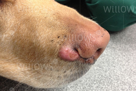
Fig 1: A mast cell tumour on the muzzle of a Labrador Retriever
Where in the Body do MCTs Occur?
Some breeds of dog are predisposed to getting mast cell tumours and tend to get MCTs more commonly in certain locations, more importantly MCTs sometimes behave in a certain way in particular breeds. For example, Pugs are renowned for large numbers of low-grade (less aggressive) tumours, and Golden Retrievers commonly get multiple tumours. Boxers with MCTs are generally younger than other breeds, and more commonly have lower-grade MCTs with a more favourable outlook. In contrast, Shar-Pei’s usually get aggressive high-grade and metastatic (spread to other sites) tumours, often at quite a young age. MCTs in Labrador Retrievers are also frequently more aggressive than in other breeds. The average age of dogs at presentation is between seven and a half years and nine years, although MCTs can occur at any age.
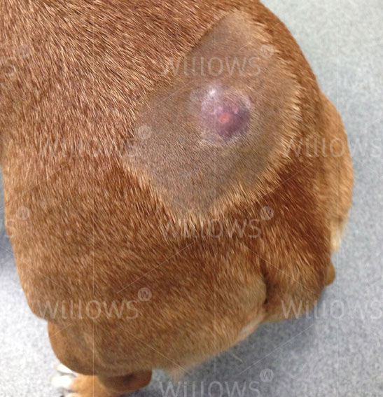
Fig 2: A mast cell tumour on a dog’s back
Other Effects of Mast Cell Tumours (Paraneoplastic syndromes)
Cancerous mast cells contain 25 to 50 times more histamine than normal mast cells. Histamine is a very inflammatory chemical, and therefore explains why some MCTs wax and wane or suddenly increase in size due to inflammation, especially after they have undergone biopsy/needle aspiration. Histamine also causes the stomach lining to produce more acid, which can result in stomach ulcers, causing signs such as vomiting or black, tarry stools (this appearance is due to the presence of digested blood coming from the ulcer).
How are Mast Cell Tumours Diagnosed?
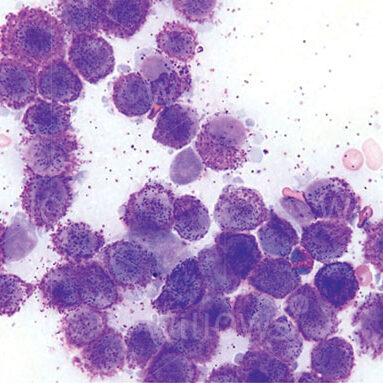
Cytology: Cytology involves looking at the cells under a microscope, the sample for this is usually obtained by ‘fine-needle aspirate’. Fine-needle aspirates of MCTs involve taking a small sample of the tumour with a thin needle. This is generally a straightforward procedure which can be done conscious and without sedation in most patients. It should be performed prior to any surgery, because a pre-operative diagnosis of MCT influences the type and extent of surgical intervention required.
Biopsy: This involves taking a larger piece of tumour tissue and sending it to a pathologist for analysis. This can be performed to help decide on the best treatment, or when the tumour has been removed to find out the grade of the tumour. The sample will be analysed under the microscope and graded to indicate how aggressive the tumour is.
Fig 3: Mast cells from an aspirate, seen under the microscope – they contain and shed typical granules. They are stained purple in this sample.
Further Investigations: ‘Staging’ of the Mast Cell Tumour
As well as performing fine needle aspiration and biopsies of the MCT to determine its grade, additional tests may be required to determine the ‘stage’ of the tumour i.e. whether or not it has already spread. These further tests can include sampling of nearby lymph nodes, chest X-rays and abdominal ultrasound scanning. The tests performed will depend on a number of factors, and these will be discussed with you by the Specialist.
Mast Cell Tumour Grade and Outlook
Low grade (grade 1) tumours and around 75% of intermediate (grade 2) tumours are cured with complete surgical excision. Unfortunately, most high grade (grade 3) tumours and around 25% of intermediate grade tumours have already spread by the time they are diagnosed (even if this spread cannot be detected on scans at the time of diagnosis). These cases benefit from chemotherapy treatment. In some dogs, further analysis of the biopsy samples is useful in determining the best management options. The tumour grade is very important in determining the appropriate therapy for dogs with MCTs.
What Treatments area Available for Mast Cell Tumours?
Surgery is the cornerstone of management of MCTs, and complete surgical removal is often curative in dogs with low or intermediate grade MCTs. To achieve a cure, in some circumstances it is however necessary to remove a significant amount of tissue surrounding the tumour must be removed to ensure that all the tumour cells are extracted. This requires a high level of Specialist surgical experience and expertise, to perform complex reconstructive surgical techniques.
If complete removal is not possible, or where the tumour appears to be more aggressive radiation therapy and chemotherapy treatments become more useful. The best treatment depends on the tumour grade, stage and other factors unique to the individual dog.
Chemotherapy can be used:
- before surgery to shrink a tumour down
- after surgery if the tumour appears more aggressive on analysis of a biopsy
- as palliative treatment if a tumour cannot be removed, has already spread or if an owner does not want to pursue surgical intervention
Fortunately, the drugs used for chemotherapy in MCTs are extremely well tolerated and most owners are very happy with their dog’s quality of life on treatment. A new group of drugs called tyrosine kinase inhibitors is also available, these block proteins (called tyrosine kinases) which are found on the surface of cancerous mast cells. They can be used where tumours cannot be surgically removed or have recurred despite previous treatments. They can have some side effects, however most dogs tolerate these drugs well.
What can I Expect if my Pet is Diagnosed with a Mast Cell Tumour?
No single factor accurately predicts the biological behaviour or response to treatment in dogs affected by MCTs. Various clinical factors can influence the outcome, such as if it has spread, potentially the breed and also the tumour location.
Tumours in the nail bed, inside the mouth, on the muzzle, in the groin area and in those sites where the skin meets mucus membranes (mucocutaneous junctions), are often correlated with a worse prognosis than those in other parts of the body, although this is not always the case.
The single most valuable factor in predicting the outcome for most patients is the grade of the tumour when assessed under a microscope (the histological grade).
What can I Expect if my Dog is Treated for a Mast Cell Tumour?
Dogs have a unique risk of developing MCTs in the skin, and they can be frustrating to manage, even for Specialist Oncologists.
Knowing what the best treatment is for an individual dog depends on knowing the grade of the MCT and whether or not it has already spread. It is important to recognise that most dogs can survive for a long time with mast cell cancer and can be cured. However, some dogs have a more aggressive type of MCT and treatment in these cases is of a more palliative nature, trying to improve patient comfort and life expectancy, but without being able to achieve a cure.
Cancer Care
Find out more
To assist owners in understanding more about Cancer Care we have put together a range of information sheets to talk you through the some of the more common conditions seen and treated by our Specialists.