Willows is one of Europe’s leading small animal Orthopaedic referral centres treating over 1000 new patients a year. Our state-of-the-art hospital is led by internationally renowned Certified Specialists committed to providing the highest standards of care.
Limb deformities are complex conditions requiring accurate assessment and experience in determining which cases require surgery and which may be managed conservatively. Our team of Orthopaedic Specialists, assisted by our Diagnostic Imaging Specialists, have extensive experience assessing animals with limb deformities and in performing the Specialist surgeries that may be required.
Why Should I Bring my Pet to Willows for Treatment of Limb Deformity?
What are Limb Deformities?
The way in which puppies and kittens limbs develop and grow is very complex. There are growth plates at the ends of the bones responsible for increasing bone length in an even manner. Occasionally growth at the plates goes wrong and deformities result.
Why do Limb Deformities develop?
Limb deformities may either be present at birth or become apparent during growth. Most growth deformities either have a genetic cause or are due to trauma. If a growth plate is injured in a young animal, for example due to a fall, it may stop growing and cause the bone to become shortened or bent.
What are the Most Common Types of Limb Deformity?
The front limbs are more commonly affected than the back limbs. This is partly because in the forearm (antebrachium) there are two bones that grow alongside each other (the radius and ulna). If one of these bones grows faster than the other, the limb will develop abnormally and become bent or twisted. Typically the paw will turn outwards.
How are Limb Deformities Investigated?
In the majority of cases a CT scan will be performed, generating 3D images that allow accurate assessment of abnormal bends (angular deformities) and twists (torsional deformities).
What Treatments are Available for Limb Deformities?
If the deformity is largely cosmetic rather than causing pain and lameness then conservative management may be recommended. Surgery is most appropriate when the deformity causes lameness or, in very young animals, to reduce the risk of a more serious problem developing.
Young dogs should be assessed as early as possible and are often treated as soon as possible to prevent progressive deformity. In adult animals, corrective surgery is elective and thus can be considered at any time if the response to conservative management is unsatisfactory.
The most common surgical procedures are:
Most commonly performed when there is a problem with one of the paired bones of the forearm. If one of the bones fails to grow normally, the growth of the paired bone may be affected. Depending on the age of the patient, the abnormal bone may be cut, this is known as an osteotomy, alternatively a section of bone may be removed which is known as an ostectomy.
What can I Expect if my Pet is Treated for Limb Deformities?
Exercise must be heavily restricted for the first six weeks after surgery. X-rays are generally obtained six weeks following surgery, with on-going rehabilitation based on both the X-ray results and an animal’s progress. Active and passive physiotherapy may be recommended to help with rehabilitation.
The outlook in dogs with growth deformities is dependent on a number of factors including the age of the dog, the degree of deformity/limb shortening, and the extent of joint involvement. Limb shortening often results in osteoarthritis that may require ongoing medical management or occasionally require ‘salvage’ surgery, such as joint replacement or arthrodesis (fusion of the joint). In cases without joint involvement the prognosis is generally good.
Case study 1

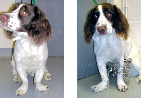
Fig 1: A Springer Spaniel with deformity of both fore limbs. The limbs are S-shaped with the paws deviating to the outside.
Fig 2: The left deformity has been corrected and the cut bones stabilised with a circular external skeletal fixator.
Case study 2

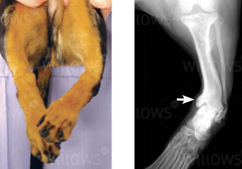
Fig 3 and 4: Photograph and X-ray of a Dachshund with severe deformity of the left hind limb. The shin bone is bent just above the ankle joint (arrow) and the paw deviates inwardly.

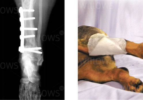
Fig 5 and 6: X-ray and photograph following surgery showing how the left tibia has been straightened and stabilised with a plate and screws.
To save this page as a PDF, click the button and make sure “Save as PDF” is selected.
Orthopaedics
Find out more
To assist owners in understanding more about Orthopaedics we have put together a range of information sheets to talk you through the some of the more common orthopaedic conditions seen and treated by our Specialists.

