What is a CT Scan?
Why Should I Bring my Pet to Willows for a CT Scan?
Willows is one of Europe’s leading small animal referral centres. Our state-of-the-art hospital is led by internationally renowned Specialists who are committed to providing the highest standards of veterinary care. Willows has some of the most advanced CT scanning equipment available in veterinary facilities in the UK, this combined with an imaging department staffed by a highly experienced team of accredited, recognised Specialists makes for an unsurpassed diagnostic imaging service. The facility and the staff are available 24 hours a day, every day of the year in order to provide the best levels of patient and client care possible.
Willows has a 64-slice CT scanner, this means that for every revolution of the X-ray tube head, up to 64 slices of imaging information are obtained. Each slice can be varied in thickness and can be as little as half a millimetre in width. Using a 64-slice scanner with a fast rotation speed, a large amount of information can be obtained in a very short space of time. A scan of a large dog’s chest for example, can be performed in as little as eight seconds. It also means that very detailed information about certain parts of the body can be obtained in a way that is unparalleled by any other imaging modality. The speed of CT means that many patients have their scans performed under sedation, rather than requiring a general anaesthetic.
What is a CT (Computed Tomography) Scan?
CT is a diagnostic tool used to look at various parts of the body, especially those made of bone (including joints), air (including lungs and the nose), and soft tissue structures- particularly those with a blood supply. CT scanning is used commonly in people, and is used extensively at Willows. It uses X-rays to produce an image, these are the same X-rays that produce the X-ray images that you may well be familiar with (called radiographs).
CT works by using a continuous beam of X-rays which spin around in a doughnut shaped support called a gantry. The ‘tube head’, which produces the X-ray beam, can spin very quickly, taking as little as 0.35 seconds with our Siemens 64 slice scanner, to completely circle around the patient. While this is happening, the patient, can be moved through the gantry by electronically moving the table top. Each revolution of the X-ray tube around the patient in the scanner builds up slices of X-ray images.
After this information is obtained, powerful computers use complex software to produce images that can be recognise and interpreted. Interpretation of the hundreds of slices of information, obtained for every patient, takes considerable time and expertise, exclusively undertaken by our team of Board-Certified Specialist Radiologists.
CT scanning has a number of advantages over both conventional X-ray radiography and other imaging techniques:
- Interpretation: CT scanning removes superimposition of overlying structures as it produces slices which are cross sections of the patient, making interpretation of complex parts of the body a lot easier.
- Abnormalities: A CT scan allows the slices to be added together electronically to give much thicker slices. In this way, we often see small abnormalities that might otherwise have been missed.
- ‘Surgeon’s Eye View: The slices from a CT scan can be electronically stacked up in any direction, allowing the radiologist to manipulate the images and effectively reconstruct the tissues. This can often help surgeons to understand disease better, as they can have a more three dimensional ‘surgeon’s eye view’ of the abnormalities.
- Speed and Detail: The 64 slice scanner is incredibly quick and has the advantage of ECG gating which can build up very detailed pictures of the heart and the tiny vessels (called coronary arteries) that support it. This is very important for imaging patients with cardiac disease that is difficult to identify on ultrasound.
CT scanning is particularly useful for looking at injected contrast agent within the body. A contrast agent is a liquid that can be injected into a patient and then watched on the CT scan as it passes around the arteries and veins, and into the organs that the blood vessels supply. The fast speed of the CT scanner enables the contract agent to be tracked.
In addition, the contrast agent has to be delivered to the body quickly so that a ‘bolus’ or discrete ‘package’ of the contrast is present, rather diffusing slowly into the body. Willows has a pressure injector linked to the CT scanner, which means that contrast can be injected into a vein very quickly while the patient is being scanned. The combination of fast scanner and pressure injector means that even small blood vessel abnormalities can be seen, and the contrast tracked as it passes through the internal organs.
What is CT used for at Willows?
CT scanning is most often used for examining noses, lungs, the contents of the abdomen, and bones. CT has revolutionised the way the veterinary profession looks at problems within complicated joints such as the elbow.
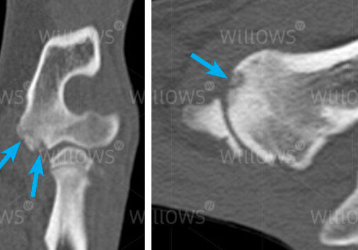
CT scans showing small erosion (arrows) on the end of the humerus (the bone of the upper limb) just above the elbow joint. This is a disease called elbow osteochondrosis, often seen in young animals. This would have been very difficult or impossible to see on conventional X-rays.
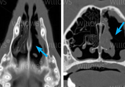
CT scans showing two views (from above and end-on) of a disease called destructive rhinitis, most commonly caused by a fungus that invades the nose. The nasal cavity on the left of each image looks normal, with lots of normal scrolled structures inside the nose called turbinates. On the other side, these delicate structures are almost entirely absent (arrows), having been destroyed by the fungus.
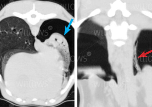
The image on the left shows a relatively normal lung (on the left of the image) and a lung that is collapsed (blue arrow). The collapsed lung is also affected by pneumonia. In the image on the right, a small straight lesion was found loose in the cavity in which the lung is housed. This was a grass seed (red arrow) which was removed surgically, without the use of CT grass seed would not have shown up on a conventional X-ray
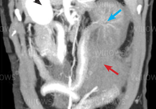
CT of a dog’s abdomen, after injecting a contrast agent, shows a normal right kidney (the white structure on the left of the image – black arrow) and a kidney on the right of the image that has a reduced blood supply (blue arrow). This dog had been hit by a car with the result that the left kidney has been torn off its blood supply – the contrast (which shows as white) is not present in the abnormal left kidney to the same degree as in the normal right kidney. The grey material (red arrow) near the affected left kidney is blood from the severed artery. The patient made a full recovery after surgery.
To save this page as a PDF, click the button and make sure “Save as PDF” is selected.
Diagnostic Imaging
Find out more
To assist owners in understanding more about Diagnostic Imaging, we have put together a range of information sheets to talk you through the some of the main areas forms of Diagnostic Imaging at Willows.