Why Should I Bring my Pet to Willows for Aspergillosis?
Willows is one of Europe’s leading small animal referral centres. Our state-of-the-art hospital is led by internationally renowned Specialists, committed to providing the highest standards of veterinary care. At Willows we have a team of Specialists in Internal Medicine, Clinical Nutrition and Diagnostic Imaging, all of whom are involved in the investigation of Internal Medicine cases.
Our state-of-the-art facilities include access to the most advanced diagnostic equipment, such as high quality ultrasound and endoscopy, to comprehensively investigate symptoms. All of which is supported by our dedicated team of Nurses and clinical support staff are available 24 hours a day, every day of the year to provide the best possible care for your pet.
What is Aspergillosis / Fungal Rhinitis?
Fungal rhinitis is an infection involving the nose and sinuses (air spaces within the skull). Fungal rhinitis in dogs is usually caused by a fungal species called Aspergillus fumigatus, known as ‘aspergillosis’ or ‘fungal rhinitis’. Aspergillus is found everywhere, particularly in soil, and so all animals and people are regularly exposed to the organisms and their spores.
Aspergillus often causes infection in the nasal cavity and the frontal sinuses of dogs. Dogs with long noses (dolichocephalic dogs) are most commonly affected, although all breeds are susceptible. Aspergillus infection in the nose can cause destruction to the normal bony scrolls (turbinates) that are present in the nose, and the fungus can form mass-like lesions called fungal plaques. It is not uncommon for the infection to spread from the nose into the frontal sinuses where it can be more difficult to treat.
In rare situations aspergillosis can affect many different systems in the body, this tends to only occur in dogs that have a problem with their immune system or that are receiving treatment to suppress the immune system.
What are the Signs of Aspergillosis / Fungal Rhinitis?
The most common signs of aspergillosis include:
- Nasal discharge; creamy or green in colour affecting one or both nostrils. Blood can sometimes be seen within the discharge
- Sneezing
- Nose bleeds (epistaxis)
- Pale discolouration of areas of the front of the nose (depigmentation of the nasal planum)
- Discomfort on examination or touch of the nose
Reverse sneezing.
How is Aspergillosis / Fungal Rhinitis Diagnosed?
Fungal rhinitis can be difficult to diagnose as the symptoms can appear very similar to other nasal diseases (e.g. nasal foreign body, nasal tumour, allergic rhinitis). A range of tests are available, however they do present some limitations:
- X-rays are very insensitive at detecting the disease within the nose
- Blood tests for antibodies to aspergillus fungus can only show that a dog may have been exposed to the fungus, and are not necessarily associated with an active infection. False negative results can also occur i.e. a dog can be affected by aspergillosis and not have any antibodies to the fungus in the blood
- Swabs taken from the nasal cavity may be grown in the laboratory and give a positive culture, but since the fungus is found so widely in the environment, its presence within the nose does not confirm an active infection
If it is suspected that a dog may be suffering from aspergillosis it is often recommended that a CT scan be performed to include the nasal cavities and the sinuses. The findings on a CT scan can often be typical for aspergillosis, enabling an initial diagnosis to be made. A CT scan will also help to rule out other conditions such as nasal tumours that can cause similar symptoms.
In some cases, a camera is used to see the inside of the nasal cavity (rhinoscopy or endoscopy) and, where necessary, to take biopsies in order to confirm the diagnosis.
In some cases, the degree of certainty given by a CT scan can be sufficient for a Specialist to recommend treatment of the fungal disease under the same anaesthetic used to perform the diagnostic tests.
What Treatments are Available for Aspergillosis / Fungal Rhinitis?
The majority of the fungal growths sit on the surface of the affected tissues and are not susceptible to treatment using injections or oral medication. As such, treatment for aspergillosis often involves the removal of these fungal plaques and/or the instillation of topical anti-fungal drugs into the nasal cavity and the frontal sinuses, performed under general anaesthesia. The treatments available at Willows vary from a minimally invasive endoscopic procedure where a camera is passed into the nose and used to thoroughly remove the fungal material. Some dogs may also require the use of anti-fungal drugs given by mouth.
What can I Expect if my Pet is Treated for Aspergillosis / Fungal Rhinitis?
The long-term out-look for dogs with aspergillosis can be very good. Depending on the treatment performed, success rates with a single treatment can approach 70-80%. A treatment plan will be specifically designed each individual pet offering the best chances of success, in some cases this can involve two procedures carried out three to four weeks apart.
A success rate of approximately 80% does mean, that for some dogs who, despite our best efforts, continue to suffer from the fungal disease, and the clinical signs do not improve as expected or that recur at a later date. In such cases, repeat or alternative treatment can be considered and it is possible that this might involve a prolonged stay with in the hospital.
Once the infection is eliminated, re-infection at a later date is uncommon. However, it cannot be ruled out and so owners are advised to be vigilant of future signs of the condition.
The damage caused by the fungal infection can make animals more susceptible to bacterial nasal infections in the future. If a nasal infection develops after successful treatment of fungal rhinitis, it is advisable to visit the a local Vet initially, as it may be possible to treated the infection in Primary Care. Persistence of nasal discharge, or the presence of blood within the discharge, is likely to increase the level of concern regarding the presence of an active fungal infection.
The outlook for patients with systemic aspergillosis is more guarded and will depend on whether we are able to establish why a dog is predisposed to this condition and whether the predisposing causes can be dealt with.

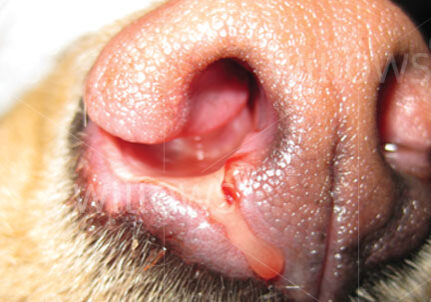
Figure 1
The front of a dog’s nose showing some blood-tinged creamy discharge from the right nostril and ulceration that can occur in some cases.


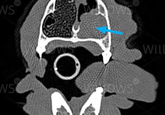
Figure 2
CT image of a cross section of a dog’s nose. The right side (seen on the left) has a fine lace-like appearance representing the scrolls of bone that are normally present. The arrow is pointing to the left side of the nasal cavity which has suffered from destruction of these turbinates and accumulation of discharge that appears the same shade of grey as the soft tissues of the dog’s head. These changes are strongly suggestive of fungal rhinitis and would be very difficult to identify on normal X-rays.
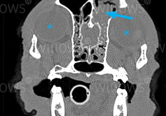
Figure 3
CT image of a cross section of a dog’s nose at the level of the frontal sinuses. The eyes can be seen as circular areas (marked with asterisks). The dog’s left frontal sinus (arrowed) has become almost completely filled with fungal growth. Air is seen as black on the scan.

To save this page as a PDF, click the button and make sure “Save as PDF” is selected.
Internal Medicine
Find out more
To assist owners in understanding more about Internal Medicine we have put together a range of information sheets to talk you through the some of the more common Medicine conditions seen and treated by our Specialists.

