What is an Ultrasound Scan?
Why Should I Bring my Pet to Willows for an Ultrasound Scan?
Willows is one of Europe’s leading small animal referral centres. Our state-of-the-art hospital is led by internationally renowned Specialists who are committed to providing the highest standards of veterinary care. Willows has very sophisticated ultrasound scanning, this combined with an imaging department staffed by a highly experienced team of accredited, recognised Specialists makes for an unsurpassed diagnostic imaging service.
The facility and the staff are available 24 hours a day, every day of the year in order to provide the best levels of patient and client care possible.
What is an Ultrasound Scan?
Ultrasound scanning is a diagnostic test used to look at various parts of the body, in particular the heart, abdomen (tummy) and other soft tissues. Ultrasound is commonly used in people (most familiarly in pregnancy), and is performed at Willows on a daily basis. Unlike X-rays, ultrasound scanning uses sound waves rather than radiation to obtain an image.
During an ultrasound scan, the patient is placed on a table, usually under sedation, and gently held. An ultrasound probe, which looks a bit like a microphone, is held against the part of the body being scanned. The probe transmits and receives ultrasound waves, and a powerful computer then analyses the waves and builds up a picture of the parts of the body which are being investigated.
Ultrasound waves do not pass through air and, because of this, the patient must be prepared carefully for the examination. The patient’s hair must be clipped in the region that is being examined, and a gel is applied to both the skin and the probe, to ensure good contact between them.
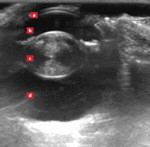
Ultrasound scanning provides superb detail in soft tissues allowing examination of parts of the body that are not easily seen on X-rays, i.e. internal structure of the heart and the eye, the contents of the abdomen and the muscles in the limbs. Conventional X-rays cannot distinguish between the outside wall and the inside cavity of hollow, fluid-filled organs, i.e. the bladder, whereas ultrasound allows these two separate parts to be identified and examined in detail.
Ultrasound scanning provides a moving ‘real time’ image, a bit like an instant movie of what the probe is ‘seeing’. This allows assessment of movement in the heart or other structures in the body, it also enables the Imaging Specialist to ‘piece together’ the images in their mind as the probe is moved around. Movement of blood in the heart and blood vessels, as well as in organs, can also be assessed. Real time imaging also allows the Imaging Specialist to take samples (biopsies) of different tissues under ultrasound guidance, where the sampling needle can be directly visualised with ultrasound in the tissues at the time the sample is being taken.
Ultrasound Scanning
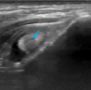
The majority of ultrasound scans performed at Willows are of the abdomen. Ultrasound provides superb detail about the internal structure of the organs, and differentiates between fluid and solid tissue. It is invaluable when there is abnormal fluid (called ‘ascites’) within the abdomen. In addition, ultrasound allows assessment of movement in real time, and so stomach and gut movement can be seen. Sampling of the different tissues and fluids within the abdomen can be performed with increased safety under ultrasound guidance, rather than being carried out ‘blind’. Below is a video of an ultrasound scan of urine entering the bladder. The urine enters the bladder via the tubes from the kidneys (the ureters) and it can be seen squirting into the bladder as the red coloured jet (on this Doppler colour-flow scan the red colour indicates a rapid flow of fluid.
To save this page as a PDF, click the button and make sure “Save as PDF” is selected.
Diagnostic Imaging
Find out more
To assist owners in understanding more about Diagnostic Imaging, we have put together a range of information sheets to talk you through the some of the main areas forms of Diagnostic Imaging at Willows.