Oral Tumours in Dogs
Why Should I Bring my Pet to Willows for Diagnosis and Treatment of Oral Tumours?
Willows is one the UKs only referral centres to have full-time Specialists in both the Medical and Surgical of tumour diagnosis and management. This, combined with a multi-disciplinary team approach to the management of pets with oral tumours, enables Willows to provide the best possible levels of care to our patients.
Our Oncology team are fully supported by Specialists in Diagnostic Imaging, Anaesthesia, Emergency and Critical Care, Clinical Nutrition and by our dedicated Interns, Nurses and Clinical support staff who provide care 24 hours a day, 365 days of the year.
What is an Oral Tumour?
Oral tumours are common in dogs and account for up to 7% of all tumours. A variety of benign and malignant tumours may be seen, along with other masses (lumps) that are not tumours. Most masses may be investigated in a similar fashion, the options for treatment and the prognosis will then be determined by the type of tumour present.
What is an Oral Tumour?
Most dogs with an oral tumour have a mass (lump) that is noticed by the owner or the Vet. Masses that are at the back of the mouth, or on the upper jaw may be more difficult to see, and therefore may only be detected when they are larger. Other signs such as reluctance to eat (particularly hard food), excessive salivation, bleeding from the mouth, smelly breath or loose teeth may also be seen.

How are Oral Tumours Diagnosed?
A diagnosis of cancer is normally made from a tissue biopsy which involves examining a piece of the tumour under a microscope. Additional tests (immunohistochemistry) may also have to be performed to determine what tumour type is present. Samples are usually also taken from the lymph nodes to look for evidence of tumour spread.
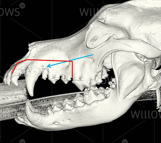
In order to assess the size and extent of the tumour and to help plan surgery, diagnostic imaging (X-rays or a CT scan) of the mouth and chest will be carried out to look for evidence of tumour spread is performed at the time of the biopsy.
Fig 1: A CT scan of the mouth to assess the size and extent of the tumour
What types of Oral Tumours can form in Dogs?
Benign Tumour and Non-Cancerous Masses
- Acanthomatous Ameloblastoma: A common benign tumour, which has the potential to grow quite large and invade the underlying bone. Tumour spread does not occur and complete surgical removal generally results in a cure.
- Peripheral Odontogenic Fibroma: A slow growing and often very firm masses that arise from the gum, usually close to the teeth. A relatively minor surgical procedure can generally be performed to remove these tumours, with good prospects for tumour control.
- Gingival Hyperplasia: A benign proliferation of the gum (gingiva), representing normal tissue that has simply grown to excess. It may be present at multiple sites in the mouth. Trimming this tissue to remove any parts that are causing difficulty when eating or affecting the health of the adjacent teeth is usually all that is needed.
- Epulis: Epulis simply means a mass on the gum that may encompass benign and malignant tumours. It is however generally reserved for benign masses, such as peripheral odontogenic fibroma and gingival hyperplasia.
Malignant Tumour
- Malignant Melanoma: Malignant melanoma is the most common oral tumour in dogs. Melanomas are often black in colour (due to melanin pigment), but some tumours may be pink (amelanotic melanoma). It may be difficult to tell the difference between this tumour type and fibrosarcoma on examination of a tissue biopsy with additional tests (e.g. immunohistochemistry) often required. Melanoma tends to grow rapidly and the surface is often ulcerated, which may lead to bleeding from the mouth and smelly breath (halitosis). Melanoma has a high rate of spread to other organs, usually the lymph nodes and then the lungs, with up to 80% of dogs developing tumour spread. Surgery to remove the primary mass, and possibly the lymph nodes, followed by administration of the therapeutic vaccine is usually used.
- Squamous Cell Carcinoma: Squamous cell carcinoma is the second most common oral tumour in dogs. This tumour arises from the lining of the mouth (mucosa) and is usually seen as a pink mass that may bleed easily. The rate of tumour spread is relatively low (up to 20%), but this may depend on the size and location of the mass, how long it has been present, and the age of the dog. This tumour may invade the bone of the jaw. Surgery to remove the mass and, if necessary, a margin of bone around the tumour is the best treatment. If the entire tumour is removed, no further therapy is required and many animals can be cured of this tumour.
- Fibrosarcoma: Fibrosarcoma is the third most common oral tumour in dogs. It arises from fibrous tissue below the lining of the mouth, and has a tendency to invade the surrounding soft tissue and bone. Under the microscope these tumours may appear relatively benign, but in the patient they usually behave in a more malignant fashion with rapid growth, invasion and tumour spread. Surgery to remove the mass and the affected bone is the best therapy. Removing the entire tumour however may be difficult as these tumours may be large or have extensive invasion into the surrounding soft tissues or bone. If all of the tumour cannot be removed, additional therapy to kill any remaining cells may be needed, such as radiotherapy or continuous low-dose (metronomic) chemotherapy.
- Osteosarcoma: Osteosarcoma is the fourth most common oral cancer and develops from the bone of the upper or lower jaw. It has the potential to spread, although this rate is relatively low. Surgical removal of the mass and the affected bone, followed by chemotherapy is the recommended therapy.
What Treatments are Available for Oral Tumours?
Surgery is used for most oral tumours, as it is the most rapid, efficient and cost-effective way of controlling the tumour in the mouth. For smaller, accessible tumours that have not spread, it offers the potential for complete cure. Many tumours will however invade the underlying bone as such successful removal of the tumour may require removal of the underlying bone. Depending on the size and location of the tumour, this may involve removal of a part or all of the lower jaw bone or a segment of the upper jaw. In the majority of dogs, this procedure is well-tolerated, and although there may be some cosmetic change, dogs continue to eat well after the procedure. Surgery may also be used to remove the local lymph nodes for diagnostic purposes to find out if there is tumour spread, or for therapeutic purposes if tumour spread to the lymph node has been diagnosed by a needle biopsy pre-operatively.
Possible complications following surgery include blood loss, wound breakdown, increased salivation, and difficulty picking up food or toys. Surgery may also result in a cosmetic change, with shortening of the jaw or a defect in the normal contour of the jaw or drift to one side.
What can I Expect if my Dog is diagnosed with an Oral Tumour?
The outlook for a patient with an oral tumour depends on the nature of the tumour, its size and its location. Surgery offers the potential to cure most dogs with benign tumours and some dogs with malignant tumours and no evidence of spread, as long as the entire tumour can be removed.
In some dogs with malignant tumours there may however be no evidence of tumour spread when the investigations are performed, yet they may still develop tumour spread in the future, which is then associated with a poor outlook. Early investigation, an accurate tissue diagnosis, appropriate diagnostic imaging and prompt therapy are key factors to improve the health and outcome of dogs with oral tumours.
To save this page as a PDF, click the button and make sure “Save as PDF” is selected.
Cancer Care
Find out more
To assist owners in understanding more about Cancer Care we have put together a range of information sheets to talk you through the some of the more common conditions seen and treated by our Specialists.